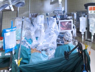


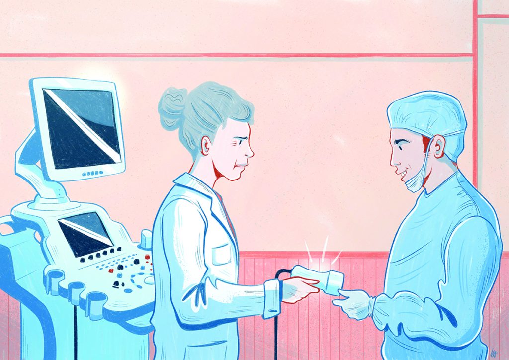
Surgeons are increasingly using ultrasound for visual guidance, both in an interoperative setting, to help ensure a clean cut around the tumour, and also to guide breast biopsies. Radiologists however are warning that performing and interpreting ultrasound scans can be trickier than is often assumed and that, in untrained hands, ultrasound could harm patient care. They worry in particular that, if surgeons get an official go-ahead to use ultrasound to help them visualise tumours, there is nothing to stop them also misusing the technique, eg to screen for or characterise lesions, which requires multimodal imaging and great expertise.
Should surgeons ever be allowed to use ultrasound? Does it make sense – and would it even be possible – to stop them using something that helps them do their job better? Is the answer to ensure any surgeon who uses ultrasound has the necessary training and experience? Or to define strict boundaries about who should do what? Or just rely on surgeons and radiologists to sort it out for themselves within the context of multidisciplinary collaboration?
Would it even make sense to operate a single approach across Europe, given that in some countries, particularly in southern Europe and the UK, the role of surgeon and radiologist have traditionally been strictly separated, while other countries, such as Germany, have a long tradition of interdisciplinary cooperation in breast cancer, and most breast surgeons are gynaecologists who learnt ultrasound as part of their obstetrics training?
The European School of Oncology invited breast surgeon Francesco Meani, Clinical Director, of the Swiss Italian Breast Centre, to debate the issue with two radiologists: Rubina Trimboli, a leading member of the European Society of Breast Imaging, EUSOBI, from the Department of Biomedical Sciences for Health, Università degli Studi di Milano, Milan; and Alexander Mundinger, from the Department of Radiology at the Niels-Stensen-Klinik, in Osnabrück, Germany, who is chairman of the executive alumni of IBUS, a non-profit organisation that has trained more than 12,000 participants in more than 120 courses since its foundation in 1991.
The debate was co-chaired by IBUS President Erkin Aribal and Alberto Costa, CEO of the European School of Oncology.
*******

Francesco Meani
To me it seems obvious why the surgeon should use ultrasound, even as a general concept. Using a device that can help you see better is a no-brainer, and should be no surprise to anybody. When you are driving a car, you prefer to drive on a sunny clear day rather than to find yourself in a bank of fog. As a surgical patient, would you like your surgeon to see the lump they are operating on, or would you prefer them to be working on you blindly? I think we need to draw a distinction between using ultrasound for the discovery and characterisation of breast lesions on the one hand, and using it to improve localisation and spatial allocation of a lesion, to guide clinical interventions, on the other. The first of these requires high-resolution ultrasound equipment, specific training and experience for imaging interpretation, and integration of different complementary modalities, and is best done by the radiologist. The second can be done by the surgeon and must be a skill acquired by both the radiologist and the surgeon. Pre- and intraoperatively there is a lot of assistance we can get from the ultrasound. We can precisely localise the lesion and evaluate the surgical margins; check the placement of the marker wire, if we used one; and check the presence and placement of the metallic clip. We can use it for intraoperative radiotherapy; and postoperatively to check the tumour bed, keep an eye out for seroma, check for abscesses and see whether we have fluid around an implant. The only two published surveys I could find on this topic indicate that surgeons are under-using this technique, probably due to lack of confidence, lack of time, limited access to ultrasound devices, and, importantly, restrictions from the radiology department or hospital. Rather than approaching this issue as a clash of interests, we should be asking what is required to give breast surgeons the capacity to use ultrasound in the best interests of patients, and we should define the boundaries we want to give the surgeon for using ultrasound, to make a good collaboration between the two parties.

Rubina Trimboli
I agree that we are a multidisciplinary team, so we have to work together in a constructive perspective for obtaining the best for our patients. But I don’t agree that there is a problem of radiology departments keeping control of the ultrasound machine. The big problem is that, if surgeons can get certification for carrying out breast ultrasound, it will be very hard to stop them from using it in the sort of discovery and diagnostic settings that we both agree are not appropriate for surgeons. We have to be responsible and only do what we are confident with and know how to do. If, as Meani says, surgeons generally lack the confidence, the time and access to up-to-date equipment to do effective ultrasound examinations, then why should they do it? Breast ultrasound is not as easy as might be assumed. There is a high proportion of human variability in determining the diagnostic result, so while the quality of the equipment clearly matters, it is one of the cases where the expertise of the examiner matters more. And with manual ultrasound examinations, quality control is difficult, because if the initial examiner overlooks an abnormality during the ultrasound examination, an image showing that abnormality will not be available for later review by a third party. We radiologists are trained to look at images. Our knowledge and understanding of ultrasound technology allows us to optimise the image to see findings that might otherwise be occult. We are also the only ones who can synthesise those findings with information that we get from other imaging modalities and clinical information of the patient. Various MRI modalities, tomosynthesis, contrast-enhanced mammography, even breast CT are all used alongside ultrasound, to build up the most reliable and accurate picture. I don’t think surgeons actually want to look at all these images, because they are more practical, and we are more… maybe philosophical. Even localisation of the lesion, one of the tasks Meani suggests can be carried out by surgeons, can be surprisingly difficult. If you ask a surgeon where is a lesion that they see in the outer quadrant on the craniocaudal view and at nipple level on the mediolateral oblique view, they will usually say it is at 3 o’clock. But actually it can be in the lower quadrant, or an inner lesion in the upper quadrant, because the oblique view is not a lateral view. And we have many cases where a surgeon or physician, sometimes even a radiologist, says ‘OK there is no lesion,’ because it is not well localised.

Alexander Mundinger
Though I’m a radiologist, I learned breast ultrasound from gynaecological colleagues, who were better at it than I was, many decades ago. This is how it started. I am now the director of several departments of radiology, and I find it difficult to interest my young colleagues in breast imaging and also breast ultrasound. They are much more interested in MRI, CT and intervention procedures. I am happy if some of them are ready to do breast ultrasound in the multidisciplinary setting. I also work at a breast centre, and our breast surgeons there do more ultrasound in daily work than the radiologists, because it doesn’t matter who does it. Some surgeons are very interested in ultrasound: they have a visual approach, including when they are in the operating theatre. Others are more kinaesthetic – they like to palpate, and they don’t really benefit from interoperative ultrasound. Everybody can learn how to do ultrasound. But of course I agree that if you are going to do it, you need to understand the physics, and know how to adjust all the variables available to get the best from the technique, which is what we teach in our IBUS courses. It’s about multimodal problem solving and making smart decisions.
Rubina Trimboli
I think it’s important to emphasise again that, however expertly used, ultrasound alone is not enough for characterising or staging a lesion. We often use several imaging modalities, and have to try to make sense of the whole picture, seen from different angles, highlighting different physical properties, and with the breast in different positions, which leads to the lesion itself changing shape. It is difficult to do and can be very time consuming. That said, I agree that surgeons should be able to use ultrasound to assist with clinical procedures. If that makes it easier, why not? And now that more and more ablative techniques are developing, maybe surgeon and radiologist have to work together. Radiologists have to know some basic knowledge in surgery and surgeons need to have some basic knowledge in radiology. The problem remains, however, once we accept that surgeons can do ultrasound, it is difficult to stop them using it in outpatient settings.
Alexander Mundinger
I’m not sure surgeons are particularly motivated to do ultrasound examinations beyond what they need to know regarding where a palpable finding is, or where a preoperative lesion lies, or intraoperatively to help guide their surgical procedure. A lot can depend on how it is reimbursed, and in most countries the reimbursement is fairly limited, or it is covered by a whole budget. It is notable that, a decade ago, our gynecologist
in Germany were very interested in per- forming ultrasound, but now that the reimbursement is included in the total budget, they use it much less.
Francesco Meani
I don’t think this should anyway be about prohibiting surgeons from using ultrasound in the outpatient clinic, or their office. The important thing is that they understand that breast diagnostics is a multimodal imaging modality and that the ultrasound examination they are doing cannot be enough. If I visit a patient, apply my ultrasound, and see something, I have to send that patient to the radiologist. That’s it. On the other hand, that shouldn’t stop me doing a core biopsy, if I think it is needed. For instance, if I have a patient on my bed on a Friday afternoon, palpating something, very afraid of what it might be, and if referring her for a biopsy would entail a week’s wait, there is no point in delaying a procedure that I am perfectly capable of doing.
Rubina Trimboli
I agree that both the radiologist and surgeon can perform a core biopsy. But in a workflow in a hospital where we are very subspecialised there are some tasks that are optimised in the radiology department. On the other hand, if you look at some of the emerging techniques… when I use a 9-gauge biopsy needle, for instance, I feel I need a surgeon! The work is not so different between the two fields. There is some overlap. Local policies should decide who does what.
Alexander Mundinger
Anybody can learn to do a biopsy. It is not difficult. In the UK even technical assistants are allowed to perform them, after adequate training. It is a question of the local system. Personally I’m happy for every biopsy my surgical colleagues perform. I and my colleagues see between 500 and 600 new cancer patients a year and an abundance of surveillance patients, and we don’t have time to do all the biopsies. It’s a question of your resources, and how you organise your workload.
Francesco Meani
I agree. There are many things that can be done by both professionals. Using ultrasound to decide which surgical incision and which kind of resection and reconstruction is going to be done is a matter for the surgeon. Diagnostic use of ultrasound is for the radiologist. But there is a grey zone, which is a field for both of us. We now need to train a new generation of breast surgeons, we need to give them adequate experience in the field and continuous practice, and we need to define indicational boundaries for the use of what I call ‘clinical interventional breast ultrasound’. That will require a structured training course that combines theoretical and practical skills, possibly with a radiology placement, exposing the surgeon to a large enough case load. Ultrasound training should be included in every curriculum for breast surgeon trainees, and radiologists should be the ones to set up and lead these programmes.
Rubina Trimboli
I agree that it is the radiologist who has to teach ultrasound to the surgeon. But radiologists can also learn from spending time with surgeons. I’ve learned many useful things in my practice from attending multidisciplinary team meetings. It is very difficult trying to discuss the implications of our findings when it’s just radiologists talking, without the involvement of a surgeon.
Alexander Mundinger
I always send my young radiologists to accompany the breast surgeon for a week in the operating theatre, to understand what is going on. If you only have the theory but no practical experience, it’s not enough. Though again you need to consider the local situation and the local interfaces between radiologists and breast surgeons. I think the training faculty at IBUS, who run ultrasound training courses, and the executive of EUSOBI, the European Society of Breast Imaging, share a similar multidisciplinary approach to education and practice, and certainly at IBUS we would welcome greater involvement by surgeons. This debate has been very helpful not least in acknowledging the reality that surgeons are increasingly using ultrasound, and that we need to ensure the training is in place and the overlap and the limits to that overlap are clear.
This is an edited version of a debate that was staged at the 20th World Congress of the Senologic International Society on Breast Healthcare, in Strasbourg, on 6 December 2018.